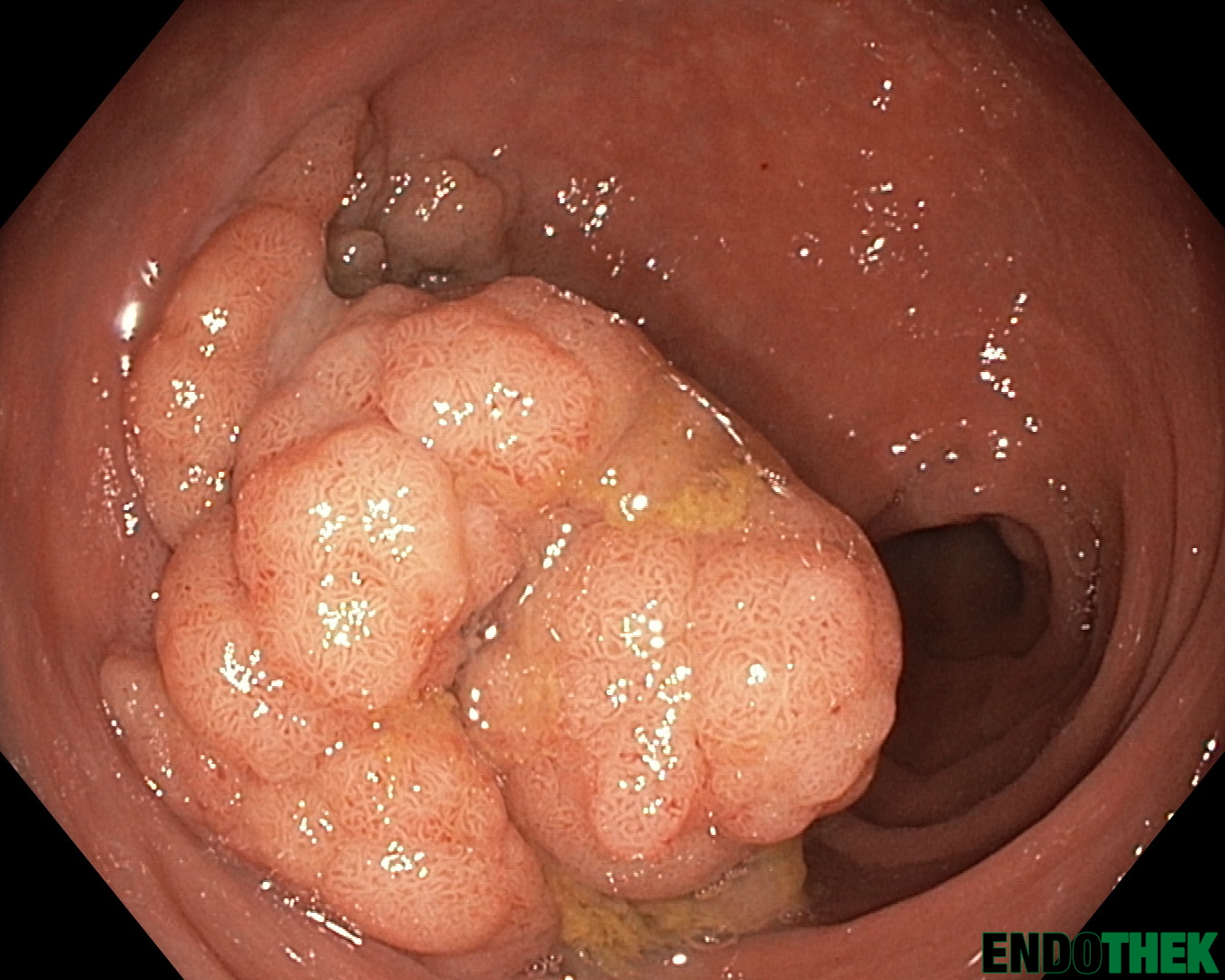
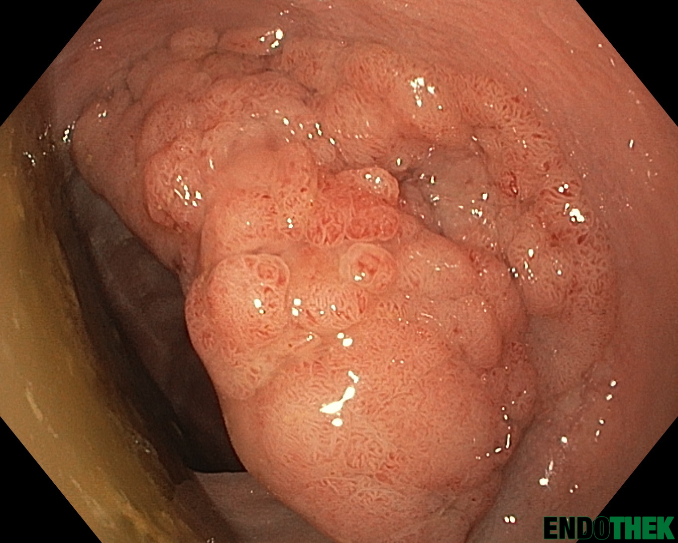
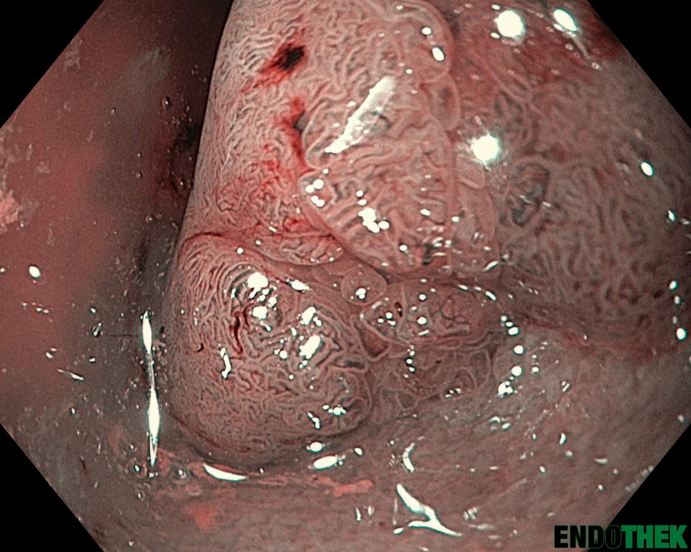
Lateral Spreading Tumor (LST) granular mixed typeLateral Spreading Tumor (LST) granular mixed type im NBILateral Spreading Tumor (LST) granular mixed type – RandzoneLateral Spreading Tumor (LST) granular mixed type – RandzoneLateral Spreading Tumor (LST) granular mixed type – Randzone im NBILateral Spreading Tumor (LST) granular mixed typeLateral Spreading Tumor (LST) granular mixed typeLateral Spreading Tumor (LST) granular mixed typeLateral Spreading Tumor (LST) granular mixed typeLateral Spreading Tumor (LST) granular mixed type im NBI, near focusLateral Spreading Tumor (LST) granular mixed type im NBI, near focusLateral Spreading Tumor (LST) granular mixed type im NBI, near focusLateral Spreading Tumor (LST) granular mixed type im NBI
Histologie: Bioptisch: Anteile eines Dickdarmschleimhautadenoms mit geringgradigen Epitheldysplasien (low grade IEN).
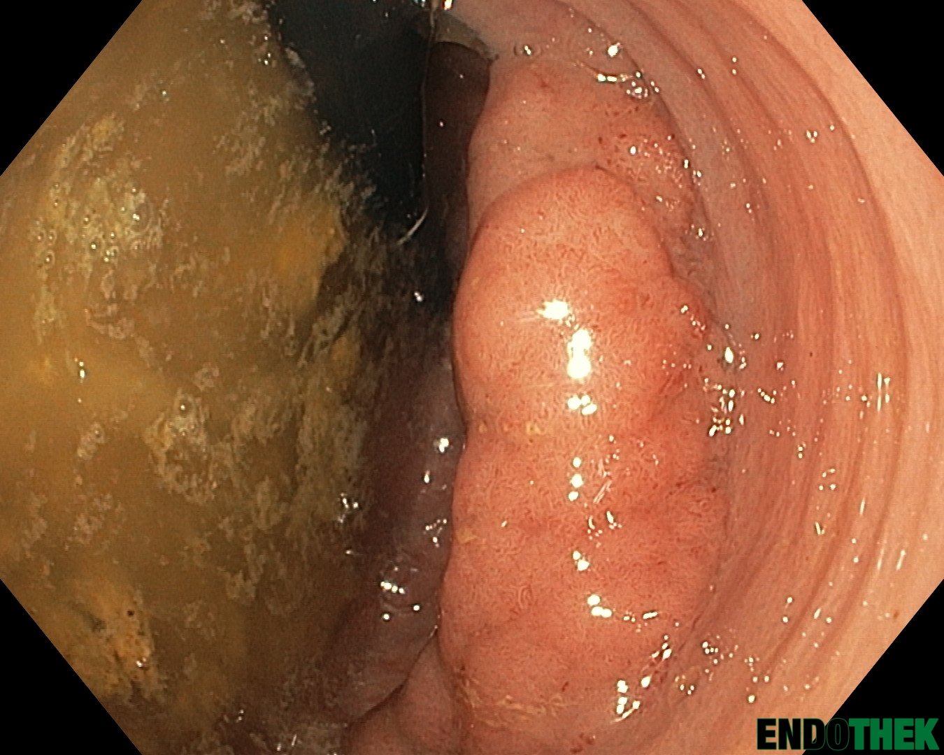
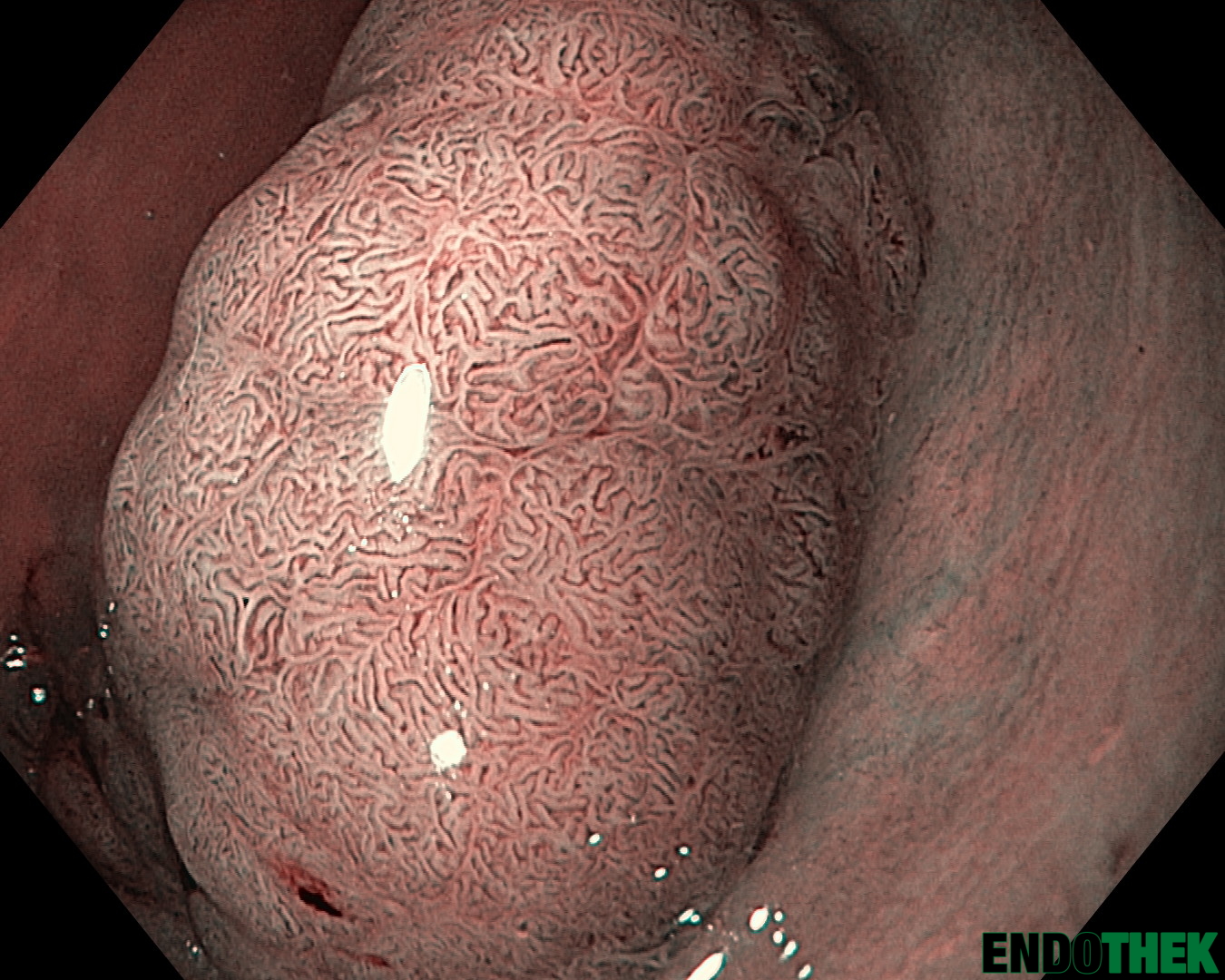
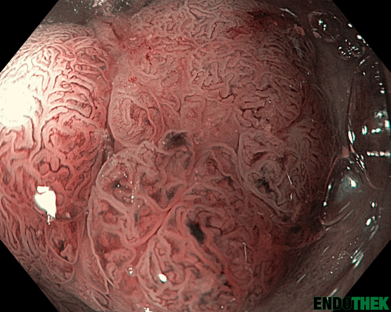
Lateral Spreading Tumor – granular mixed type im Rektum
LST granular mixed type im Rektum (NBI) near focusLST granular mixed type im RektumLST granular mixed type im Rektum (NBI)LST granular mixed type im Rektum
Histologie: (bioptisch) Zahlreiche kleine oberflächliche Fragmente eines tubulovillösen Adenoms der Dickdarmschleimhaut mit geringgradiger Epitheldysplasie (intraepithelialer Neoplasie). Ob ein anderer Stelle bereits weiter fortgeschrittene neoplastische Veränderungen vorliegen, kann naturgemäß nicht beurteilt werden.
Histologie: (in toto nach ESD) Endoskopisches Mucosadissektat (ESD-Präparat) der Dickdarmschleimhaut mit einem tubulovillösem Adenom (gering- und hochgradige intraepitheliale Neoplasie. Kein Karzinom. Das Adenom erreicht fokal die von Ihnen angebrachte grüne Tusche (lateralen) Resektionsrand. In diesem Bereich eine geringgradige (keine hochgradige) intraepitheliale Neoplasie nachweisbar
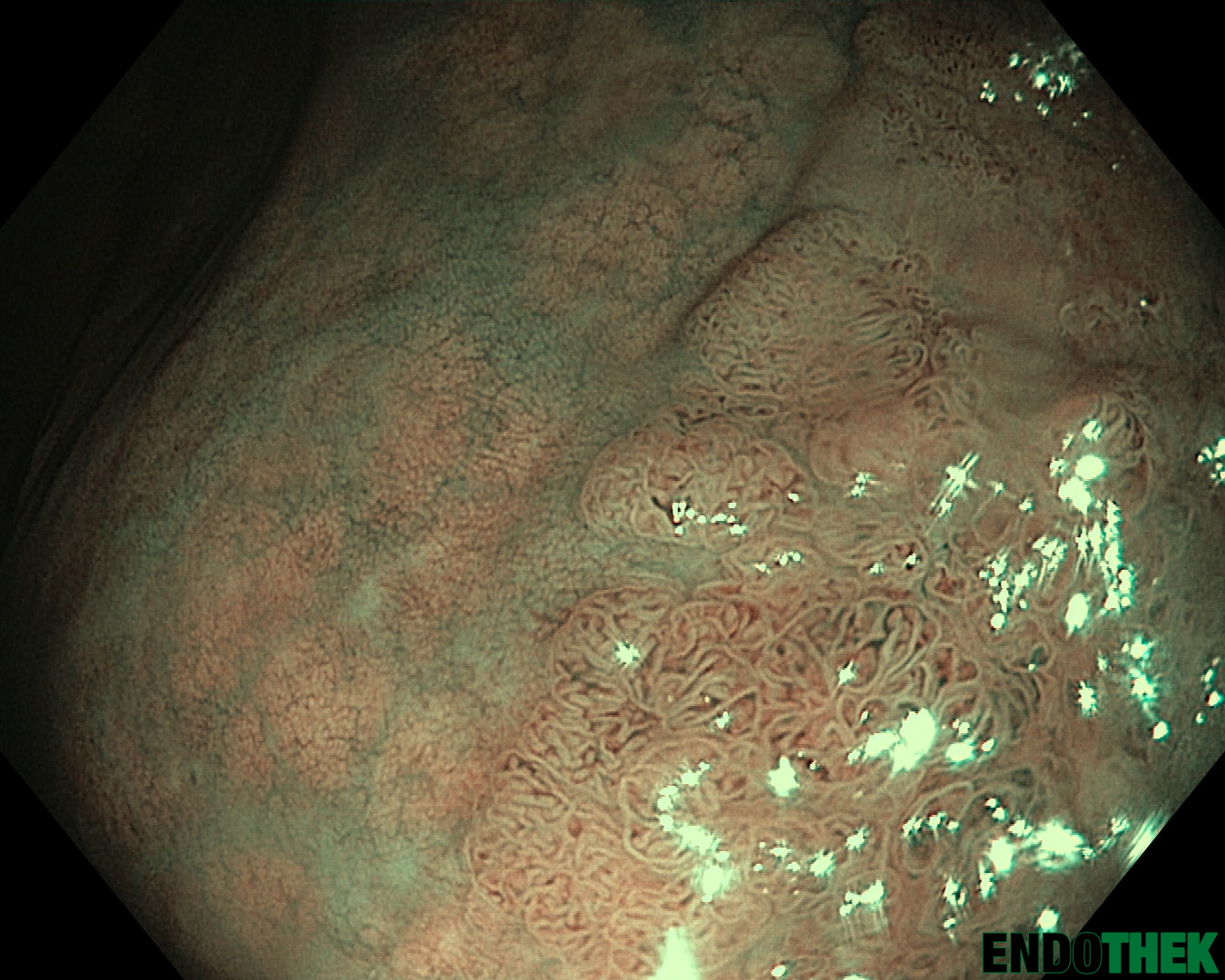
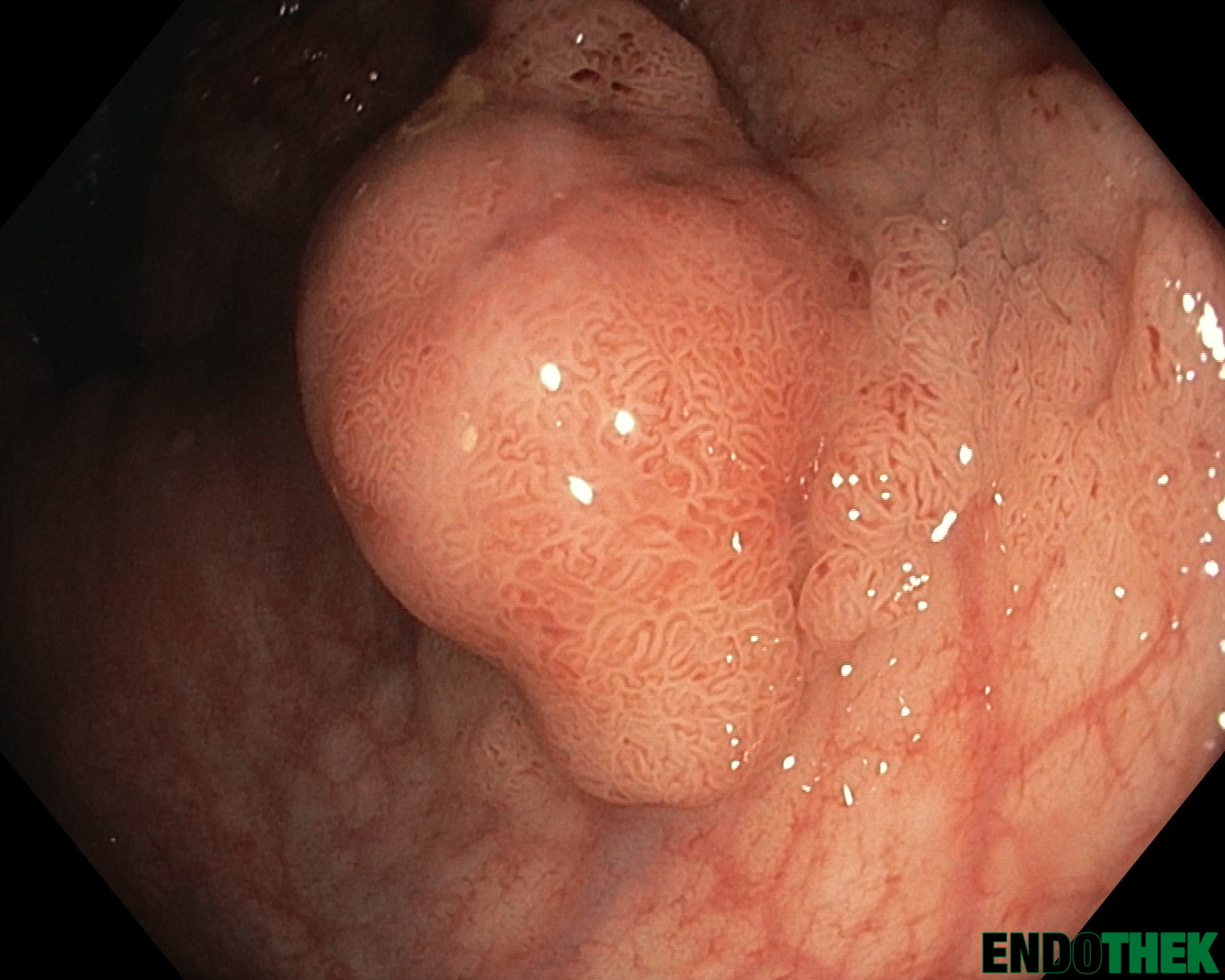
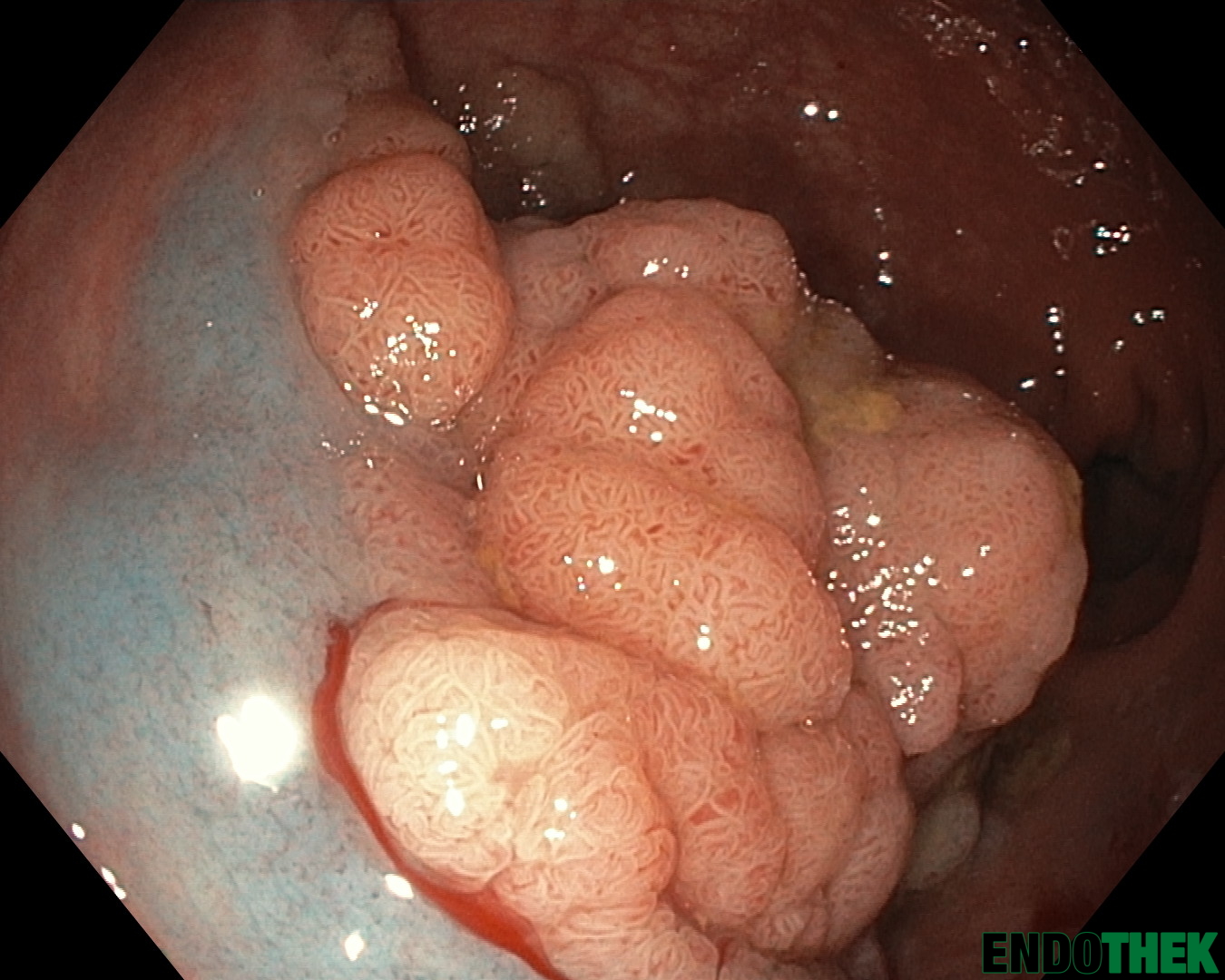
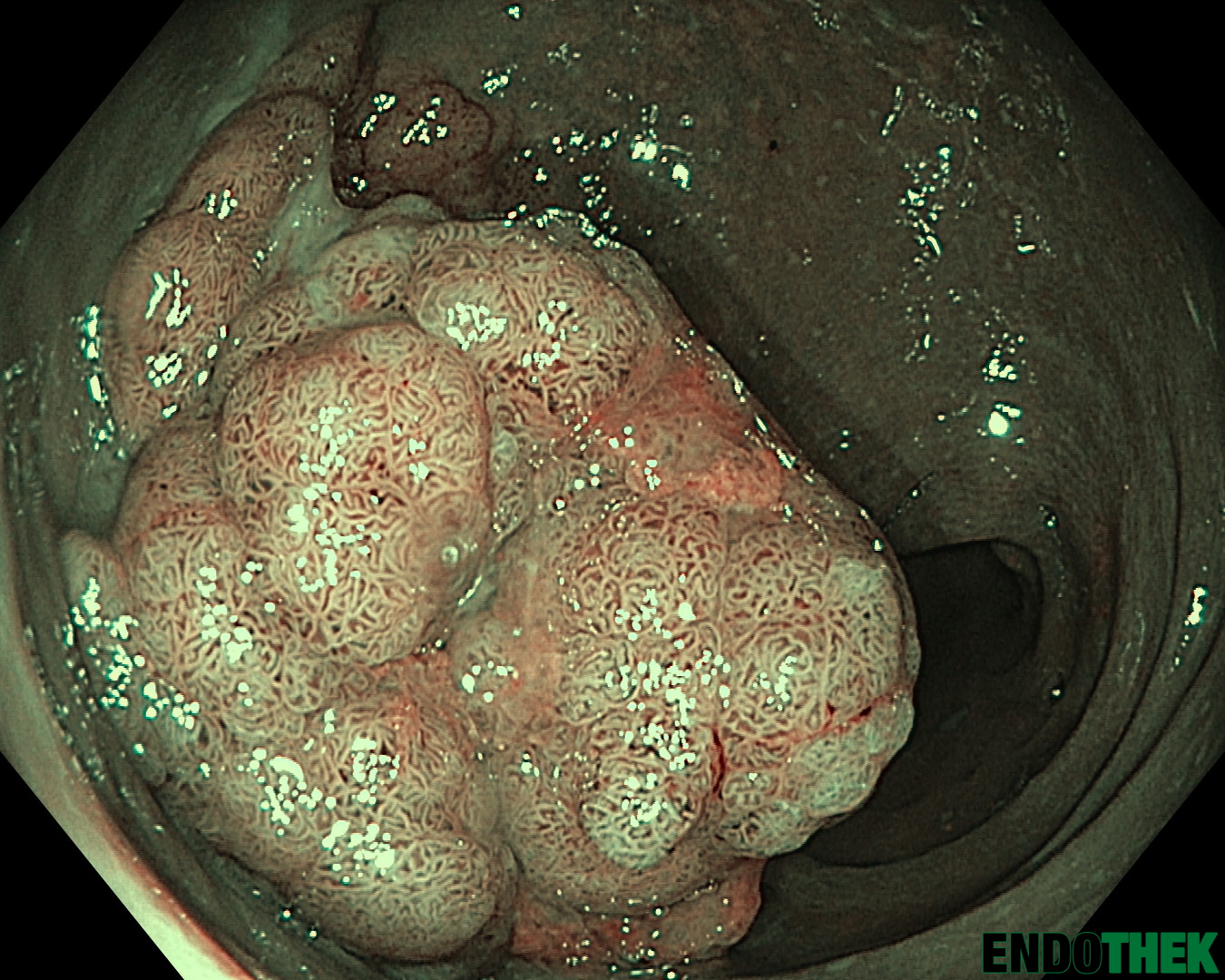
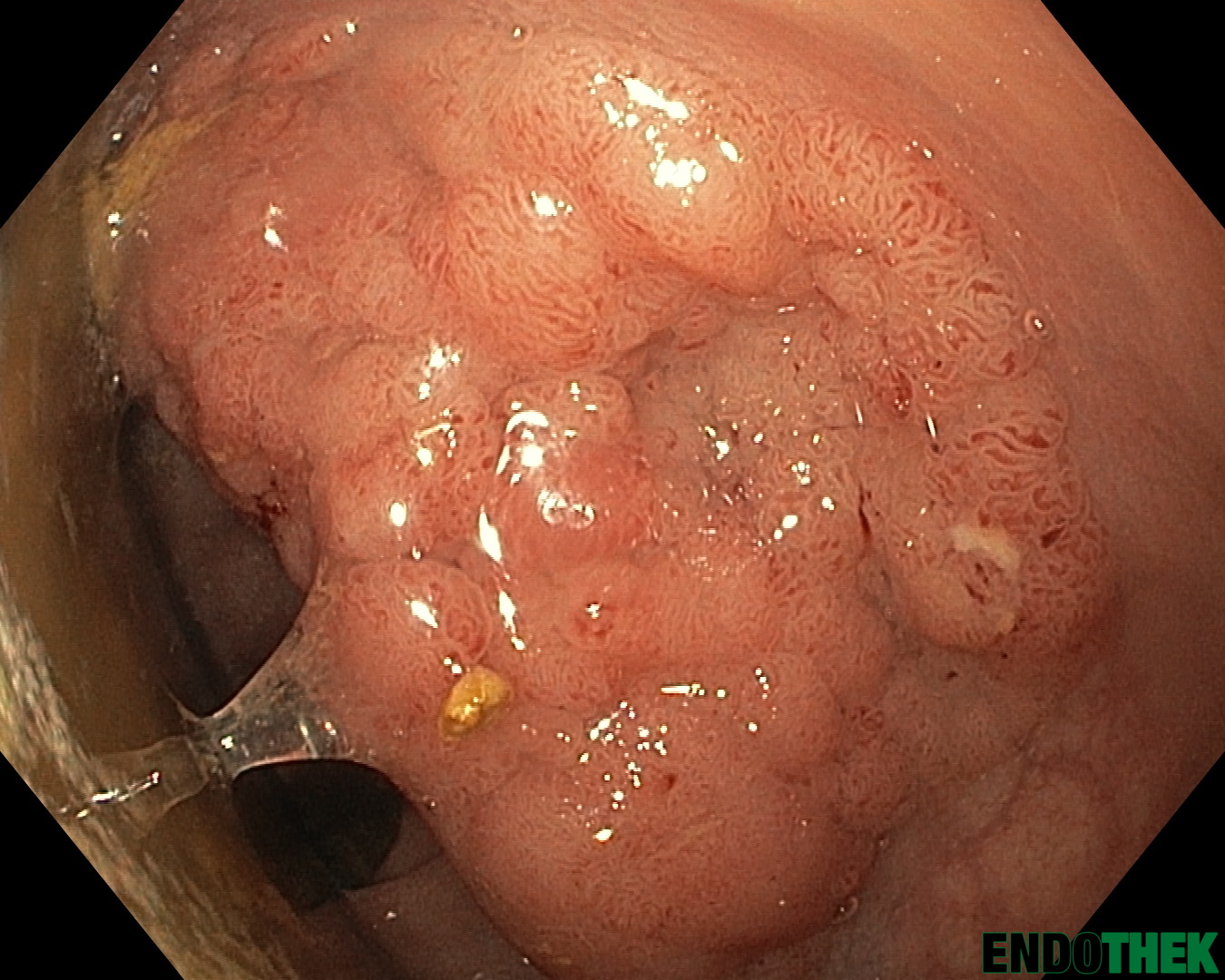
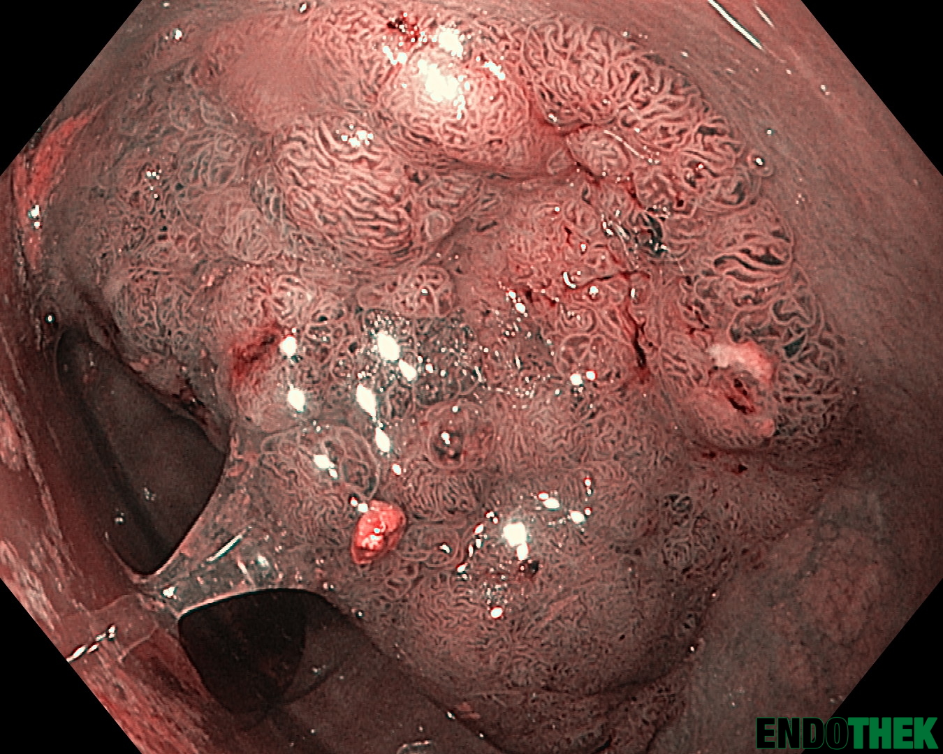
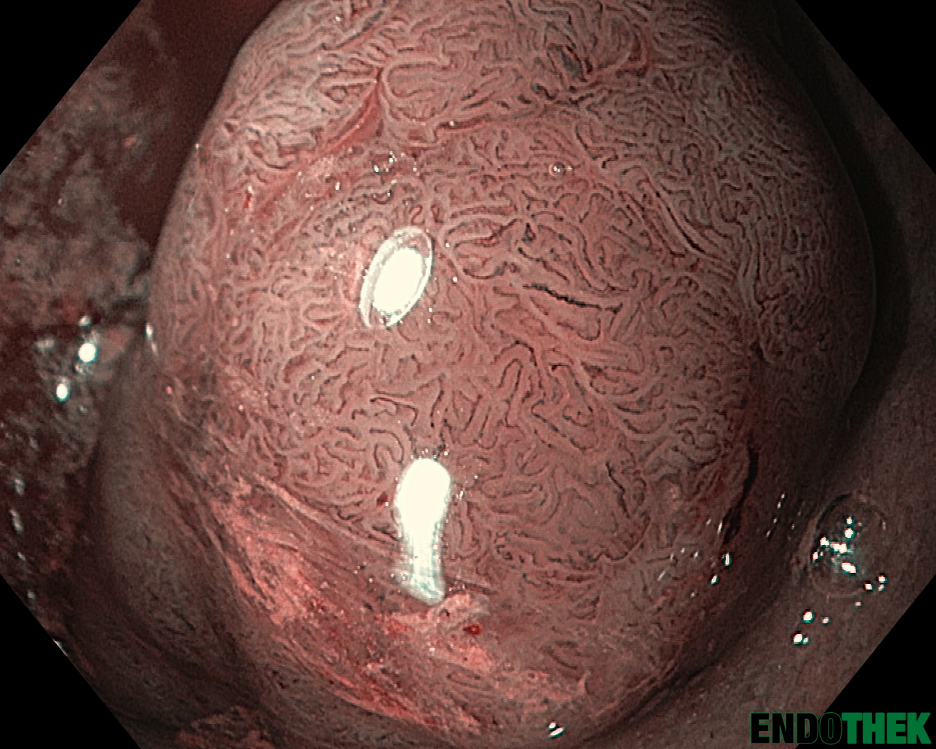
Lateral sprading Tumor – 5cm ab der Linea dentata JNET 2A/2B
LST granular type – JNET 2A/2BLST granular type – JNET 2A/2BLST granular type – JNET 2A/2B (NBI)LST granular type – JNET 2A/2B (NBI, near focus)LST granular type – JNET 2A/2B (NBI, near focus)LST granular type – JNET 2A/2BLST granular type – JNET 2A/2BLST granular type – JNET 2A/2B (NBI, near focus)LST granular type – JNET 2A/2B (NBI, near focus)LST granular type – JNET 2A/2BLST granular type – JNET 2A/2B (NBI, near focus)LST granular type – JNET 2A/2B (NBI, near focus)LST granular type – JNET 2A/2B (NB)
Histologie:
Biopsie: Mehrere Biopsieteilchen aus zumindest einem tubulovillösen Adenom (intraepitheliale Neoplasie) der Dickdarmschleimhaut mit low grade-Dysplasie und high grade-Dysplasie. Im vorhandenen, sehr oberflächlich entnommenen Gewebsmaterial, kann histopathologisch eine Stromainvasion nicht belegt werden, naturgemäß jedoch auch nicht ausgeschlossen werden.
To provide the best experiences, we use technologies like cookies to store and/or access device information. Consenting to these technologies will allow us to process data such as browsing behavior or unique IDs on this site. Not consenting or withdrawing consent, may adversely affect certain features and functions.
Functional
Always active
The technical storage or access is strictly necessary for the legitimate purpose of enabling the use of a specific service explicitly requested by the subscriber or user, or for the sole purpose of carrying out the transmission of a communication over an electronic communications network.
Preferences
The technical storage or access is necessary for the legitimate purpose of storing preferences that are not requested by the subscriber or user.
Statistics
The technical storage or access that is used exclusively for statistical purposes.The technical storage or access that is used exclusively for anonymous statistical purposes. Without a subpoena, voluntary compliance on the part of your Internet Service Provider, or additional records from a third party, information stored or retrieved for this purpose alone cannot usually be used to identify you.
Marketing
The technical storage or access is required to create user profiles to send advertising, or to track the user on a website or across several websites for similar marketing purposes.